The 3D model of the root canal system of the sample presented in Fig.
Download scientific diagram | The 3D model of the root canal system of the sample presented in Fig. 2. The additional canal is marked by arrow from publication: Root Canal System Analysis with a Group of First Permanent Molars of Upper and Lower Jaw | A progressive bacteria invasion on tooth tissues leads to pulp inflammation, microabscesses of the pulp, destruction and in consequence inflammation of periapical tissues. Therefore the aim of endodontic treatment is three dimensional debridement of a root canal from the vent | Root Canal, Molar and Endodontics | ResearchGate, the professional network for scientists.

Root Canal Anatomy of Maxillary and Mandibular Teeth

3-D image shows the enamel, dentin, and root canal with surface
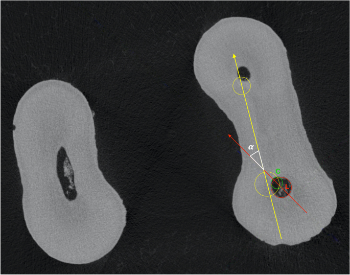
Micro-computed tomographic evaluation of endodontic ledge position

The 3D model of the root canal system of the sample presented in

Root Canal Anatomy
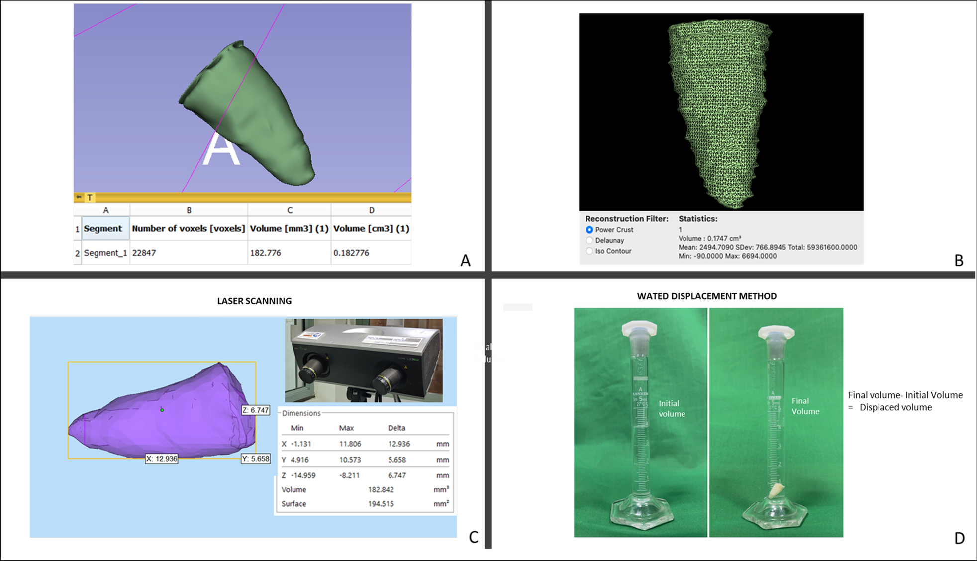
Three-dimensional semi-automated volumetric assessment of the pulp

Primary human teeth and their root canal systems - Cleghorn - 2010

Schematic diagrams showing the root canal configuration according

Root Canal Anatomy

Root canal type according to Vertucci's classification [10]. Type

Aetiology, incidence and morphology of the C‐shaped root canal

Aetiology, incidence and morphology of the C‐shaped root canal

3D models of mandibular premolars showing A: RG with different